 |
|||||||||||||||||||
|
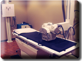
Sports MedicineSports Medicine encompasses care for patients with recent sports injuries, dislocated knees, shoulders, joints, and acute fractures, and other similar acute orthopedic injuries. Full on site xrays and physical therapy services are available. Acute injuries and Orthopaedic Prompt CarePrompt care service is for patients who have suffered an acute orthopedic injury on the field, or as a weekend warrior who are in need of care. Local Sports Team and Past Pro Sports Team ExperienceDr. Sotta is passionately involved in local high school community sports teams in the Lake Oswego, Tigard, Portland area.He was the assistant team physician for the following professional sports teams:
Team physician for many high school teams since 1980. Dr. Sotta has been in private practice in Orthopaedic Surgery and Sports Medicine in the Portland area since 1998. RehabilitationReconstructive Surgery
Orthopaedic SurgeryArthroscopic SurgeryArthroscopy is both a diagnostic and corrective procedure used in the treatment of orthopaedic conditions. The approach is minimally invasive in nature, and requires that only small incisions be made to the skin surface. Through these incisions, a small camera and tools are inserted into the joint's interior, allowing the surgeon and team to observe surgical efforts via a live broadcast feed. While an arthroscopic approach is frequently used to determine the extent and severity of damage present. Arthroscopy involves a small camera is used to view (-scopy) a joint (-arthro). This process lets the orthopedic surgeon see the inside of the knee. This facilitates accurate diagnosis and treatment of knee, shoulder, and joint problems. Advances in high-resolution cameras and improved arthroscopes have made this procedure extremely effective in accurately diagnosing and treating joint disorders. Conditions which can be treated using Athroscopic SurgeryCases of both wear-based inflammation and injury-associated damage may be subject to arthroscopic intervention. Frequently treated conditions include, but are not limited to:
Elbow & Upper Extremity
Spine, Neck & Shoulders
This is the spine neck and shoulder
information section here. Foot, Ankle & Knee SurgeryThe Anatomy Of The KneeThere are three bones that make up the knee. They are the femur, tibia, and patella. The area where these three bones meet is covered with articular cartilage. This is a smooth cushioning substance that allows the bones to move with ease.The rest of the surfaces of the knee are covered in a smooth, thin tissue liner known as synovial membrane. Synovial membrane creates a lubricating fluid that reduces friction in the knee to almost nothing as long as the knee is healthy and everything is functioning properly. The largest joint in the human body is the knee. It is used in most activities of daily living. As mentioned previously, it is made up of three bones. The femur is the lower end of the thigh bone. It rotates on the upper end of the tibia, or shin bone. The patella is the kneecap. It slides on a groove located at the end of the femur. The femur and tibia are attached by large ligaments that give stability. The large thigh muscle adds strength to the knee structure. Problems With the KneeIdeally, all the components of the knee will perform together perfectly. However, normal wear and tear caused by work and sports, along with damage caused by aging, weakening tissues, arthritis and injury can cause pain and a loss in functioning ability. Here are some problems that can be diagnosed and treated by the use of arthroscopy:
Sprains, Strains, & Dislocations
This section is courtesy of the American Academy of Orthopaedic Surgeons
Sometimes called a slipped or ruptured disk, a herniated disk most often occurs in your lower back. It is one of the most common causes of low back pain, as well as leg pain (sciatica). Between 60 and 80 percent of people will experience low back pain at some point in their lives. A high percentage of people will have low back pain caused by a herniated disk. Although a herniated disk can sometimes be very painful, most people feel much better with just a few months of nonsurgical treatment. Your spine is made up of 24 bones, called vertebrae, that are stacked on top of one another. These bones connect to create a canal that protects the spinal cord. Five vertebrae make up the lower back. This area is called your lumbar spine.  Parts of the lumbar spine.
Other parts of your spine include: Spinal cord and nerves. These "electrical cables" travel through the spinal canal carrying messages between your brain and muscles. Intervertebral disks. In between your vertebrae are flexible intervertebral disks. They act as shock absorbers when your walk or run. Intervertebral disks are flat and round, and about a half inch thick. They are made up of two components:  Healthy intervertebral disk (cross-section view).
A disk begins to herniate when its jelly-like nucleus pushes against its outer ring due to wear and tear or a sudden injury. This pressure against the outer ring causes lower back pain.  A herniated disk (side view and cross-section).
If the disk is very worn or injured, the jelly-like center may squeeze all the way through. Once the nucleus breaks — or herniates — through the outer ring, pain in the lower back improves. Sciatic leg pain, however, increases. This is because the jelly-like material inflames the spinal nerves. It may also put pressure on these sensitive spinal nerves, causing pain, numbness, or weakness in one or both legs. In many cases, a herniated disk is related to the natural aging of your spine. In children and young adults, disks have a high water content. As we get older, our disks begin to dry out and weaken. The disks begin to shrink and the spaces between the vertebrae get narrower. This normal aging process is called disk degeneration. Risk FactorsIn addition to the gradual wear and tear that comes with aging, other factors can increase the likelihood of a herniated disk. Knowing what puts you at risk for a herniated disk can help you prevent further problems. Gender. Men between the ages of 30 and 50 are most likely to have a herniated disk. Improper lifting. Using your back muscles to lift heavy objects, instead of your legs, can cause a herniated disk. Twisting while you lift can also make your back vulnerable. Lifting with your legs, not your back, can protect your spine. Weight. Being overweight puts added stress on the disks in your lower back. Repetitive activities that strain your spine. Many jobs are physically demanding. Some require constant lifting, pulling, bending, or twisting. Using safe lifting and movement techniques can help protect your back. Frequent driving. Staying seated for long periods, plus the vibration from the car engine, can put pressure on your spine and disks. Sedentary lifestyle. Regular exercise is important in preventing many medical conditions, including a herniated disk. Smoking. It is believed that smoking lessens oxygen supply to the disk and causes more rapid degeneration. For most people with a herniated disk, low back pain is the initial symptom. This pain may last for a few days, then improve. It is often followed by the eventual onset of leg pain, numbness, or weakness. This leg pain typically involves the leg below the knee, and foot and ankle. It is described as moving from the back or buttock down the leg into the foot. Symptoms may be one or all of the following:
To determine whether you have a herniated lumbar disk, your doctor will ask you for a complete medical history and conduct a physical examination. The diagnosis can be confirmed by a magnetic resonance imaging (MRI) scan. Medical History and Physical ExaminationAfter discussing your symptoms and medical history, your doctor will examine your spine. During the physical examination, your doctor may conduct the following tests to help determine the cause of your low back pain. Neurological examination. A physical examination should include a neurological examination to detect weakness or sensory loss. To test muscle weakness, your doctor will assess how you walk on your heels and toes. Your thigh strength may also be tested. Your doctor can detect any loss of sensation by checking whether you are numb to light touch in the leg and foot.  Illustration showing straight leg lift test.
Straight leg raise (SLR) test. This test is a very accurate predictor of a disk herniation in patients under the age of 35. In this test, you lie on your back and your doctor lifts your affected leg. Your knee stays straight. If you feel pain down your leg and below the knee, you test positive for a herniated disk. Imaging TestsTo help confirm a diagnosis of herniated disk, your doctor may recommend a magnetic resonance imaging (MRI) scan. This scan can create clear images of soft tissues like intervertebral disks. In the majority of cases, a herniated lumbar disk will slowly improve within 6 to 8 weeks. By 3 to 4 months, most patients are free of symptoms. Nonsurgical TreatmentUnless there are neurological deficits — muscle weakness, difficulty walking — or cauda equina syndrome, conservative care is the first course of treatment. It is not clear, however, that nonsurgical care is any better than letting the condition resolve on its own. Common nonsurgical measures include: Rest. Usually 1-2 days of bed rest will calm severe back pain. Do not stay off your feet for longer, though. Take rest breaks throughout the day, but avoid sitting for long periods of time. Make all your movements slow and controlled. Change your daily activities so that you avoid movements that can cause further pain, especially bending forward and lifting. Anti-inflammatory medications. Medicines like ibuprofen or naproxen may relieve pain. Physical therapy. Specific exercises can strengthen your lower back and abdominal muscles. Epidural steroid injection. In this procedure, steroids are injected into your back to reduce local inflammation. Of the above measures, only epidural injections have been proven effective at reducing symptoms. There is good evidence that epidural injections can be successful in 42-56% of patients who have not been helped by 6 weeks or more of other nonsurgical care. Overall, the most effective nonsurgical care for lumbar herniated disk includes observation and an epidural steroid injection for short-term pain relief. Surgical TreatmentA small percentage of patients with lumbar disk herniations require surgery. This includes the urgent surgeries for people with neurological deficits or cauda equina syndrome. Surgery for lumbar herniated disk is controversial. Research shows that patients 2 years post-surgery have the same results as patients treated nonsurgically. Patients with significant sciatica who have surgery, however, have better and more rapid pain relief. Surgery resolves symptoms faster for those with motor weakness or numbness, as well. Procedure. The most common surgical procedure for a herniated disk in the lower back is a lumbar microdiskectomy. Microdisketomy involves removing the herniated part of the disk and any fragments that are putting pressure on the spinal nerve. Rehabilitation. Most patients do not require formal physical therapy after surgery. A simple walking program 30 minutes each day, along with flexibility exercises for the back and legs, can be done as a home program. | ||||||||||||||||||


